Saturday, February 28, 2009

NOOPY

Asbcesses are sometimes really are pain in the ass. What is more annoying is the development of abscesses at the IV site. This actually, can be prevented by aseptic technique, monitoring and changing of the IV site every now and then.


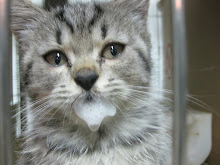
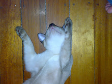
This is noopy and she has an abscessed arm. Abscessation are actually tricky to manage and may or may not require a few days of confinement and monitoring. As for the owners it was ok for them for noopy to stay awhile during the treatment.

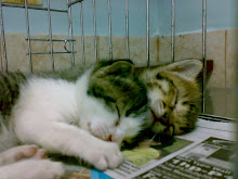
Day 1: severe abscessation. The area was flushed with NSS, PenG, Povidone lodine with table sugar and nitrofufazone was added prior to bandaging…

Daily, the wound was cleaned, unfortunately, was not documented…But, there was improvement. We noticed that the area became clearer with granulation tissue formation. There was no noticed foul odor versus the first day and the wound became smaller


Day 7: the wound is clean, smaller and appeared to have healthy granulation tissue. With formation of granulation tissue, flushing of the wound is important. Rubbing the area even with a sterile gauze that can remove the newly formed tissues should be avoided.

Day 8: the wound was left open to dry and fortunately, noopy went home today without the need for bandage.


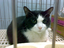
Noopy visited the clinic for vaccination update (which was again overdue!!! What’s the use of appointment slips and recommendations?!) and the arms that was affected was already clear, with hairs regrown and no evidence of previous abscess formation.


Going back to the case, why did I use sugar in the wound dressing?

Actually, I learned the technique when my father’s bed sores were being treated by his surgeon prior to grafting. He explained that the bacteria present will ferment the sugars first before it will digest the protein of the skin, and with the presence of antibiotics and povidone iodine, the microbes will die. Further research also revealed that sugars tend to promote better granulation tissue formation, decrease edema formation, attraction of macrophages, source of cellular energy and for necrotic slough . What’s more encouraging is that it is readily available and inexpensive. Aside from table sugar, dextrose powder and honey may also be used.


GOOPS


Goops was presented at the clinic due to swelling observed at the right side of the face (at about the area of the submandibular-parotid areas) just ventral to the ear to the ears. Previous history revealed bullying by other dogs.


Initial check-up revealed several scratches at the face but without deep wounds, with dark-brown, grainy otic discharge of minimal amount and an attached, firm swelling of the submandibular/parotid areas about 2 inches in diameter.
Ear swab revealed earmites.
Aspiration of the swelling revealed blood. Thus the swelling must be hematoma. The area was aspirated and the swelling subsided. Medications and compress were recommended.


A few days later, goops was again brought to the clinic due to the same problem, this time, bigger. Upon inquiry, the owner was not able to follow recommendations.

This time, drainage thru penrose drain, was placed and the area was bandaged, with Elizabethan collar placed to avoid scratching and removal of the bandages. Medications, wound cleaning and warm compress was again recommended for strict compliance.


A few days later, the owner reported back that the swelling already subsided without any complications and medications were given diligently.


By the way, this case earned us a family pizza from domino’s and a roasted chicken from andok’s… thanks to the client.


Wednesday, February 11, 2009

Fortune!

Meet Fortune! The cute, sweet and friendly Pomeranian. Cute, Yeah! Sweet and Friendly?! Nah! I strongly disagree, and I think most us think so too. Hehehe. Well, actually if you're gonna pet him and take him outside the cage especially if you're new, he will like you, and act friends with you. But when he sees someone he likes more especially if you've already put him down on the floor or on the table, he'll growl or worse bite you if ever you'd want to hold him. Maybe that's why he stayed here boarding longer than he could have stayed at home with his master. Poor Fortune... if you could just lose the biting.

Tree Kangaroo

I didn't actually know tree kangaroos exist. I haven't seen one until I saw one in the clinic, here in the Metropolis! Well actually, the owners imported her (I did not ask from where) and designed a room in their house similar to its habitat. But it is still important to leave them alone in their natural habitat and to protect the natural place where they dwell is the most valuable and effective form of keeping them alive and safe.

Apparently I touched its coat (just like a stuffed toy) only a little coarse. And it had a pouch too on its abdomen! With its long claws, it may have scratched me if it was feeling better already, told the owner. Huah!!!


Tuesday, February 10, 2009

Computed Tomography (CT) Scan is one of very important diagnostic imaging procedure nowadays used in humans. It is now also used in animals. It uses x-ray images to create a three-dimensional image of the body and its inside parts and organs.

There are so many uses for CT scan and one of the important use for this is for tumor and or cancer detection of the vital organ , imaging of compound fractures, evaluation of a spinal injury and many others.


I was able to observe several scanning appointments during my stay at AI AMC. In this case, the Dachshund is being evaluated for spinal injury which caused its hildlimb paralysis.

Usually after evaluation of cases like this, then you may be able to tell the possible treatment and therapy that may be employed and the probable outcome of the case.

Capillary hemangioma excision

When I first saw this dog, the clinic (AI Animal Medical Center) smelled like a used shoe which was stored in a closet while still wet. Basically, by his smell, you will know he is nearby. Why? Because of the ears. Apparently the ear has a bleeding wound, dirty,wet and smelly. Biopsy was taken and was found to be a capillary hermangioma.
A hemangioma is a benign (non-cancerous) vascular tumor (versus hemangiosarcoma) which usually affects older dogs. Basically it presents as a solitary slow growing nodule which may be left alone unless it causes a problem. In this dog, it is ulcerated and is non healing with possibility of getting infected thus its only cure is its excision. In this case, an ear capillary hemangioma excision was done.
This is the retriever while prepared for the surgery. Preanesthetics are given through the intravenous catheter.
After induction of anesthesia, the area is clipped, cleaned and disinfected thoroughly. Notice that the ear is soiled due to mixture of exudates and dirt.
The tumor is incised and in this photo, the wound is being cautherized to prevent further bleeding.

This is the ear after excision. Notice the location of the already removed tumor.

After the removal, the wound is carefully stitched to close the skin but not obscuring the ear canal.

After surgery, there was no follow up visit. Most likely, the owner of the dog (who was a medical doctor) removed the stitches at home.

Legg Calve Perthes Disease

About more than two months ago, Terri, a Jack Russel Terrier, underwent femoral head ostectomy (removal of the femoral head) of the right hip. This February, during the orthopedic workshop, the left femoral head will also be removed.
Why is that?
It is because Terri has been diagnosed with Legg Calve Perthes Disease (Avascular, Aseptic developmental osteonecrosis of the femoral head and neck).
What?! Basic Terms Please!
This is a genetic disease of small breeds where there is decreased blood supply (avascular) to the femoral head and neck causing that part of the body tho die (osteonecrosis) aseptically (no infection). The death of the area causes the irregularly shaped femoral head with new bone spicules growing. These bone spicules or fragments causes pain and limping as it bumps and grind with the acetabulum (socket of the hip where the femoral head fits).
This is Terri before the surgery, after induction of anesthesia.

This radiograph was taken a day before the second surgery, notice that the right femoral head (your left) is already gone (operated November 2008). Now, its time for the left femoral head to be removed.
Femoral head ostectomy will prevent pain and limping experienced by the dog due to the contact of new irregular bone spicules with the acetabulum. And since JRT's are fairly muscular dogs, they are able to gain good gait a few weeks after surgery provided adequate care and management is given.

Monday, February 2, 2009


See this Corgi undergo his regular swimming therapy with his doctor, Dr. Matsumoto

Swimming Therapy

See this Corgi undergo his regular swimming therapy with his veterinarian, Dr. Matsumoto.

As an good form of therapy and exercise, swimming stimulates blood circulation and strengthens the muscle and joints without much impact to the bones and associated joints. It is also a good cardio work out.

Water Treadmill

Some may find this amusing. Some won't believe what this would do to your pets, but yes, this is specially designed for your pets that need therapy. This may help your pets to be able to walk again. Though it is entirely a case to case basis if your pet can ever walk again, i think you just have to ask about this with your veterinarian.

This technology is usually employed in countries that are able to afford this facility like Japan. Maybe in the near future, this will be present here in the Philippines. for now, we can still do with the swimming therapy, ultrasound therapy, guided/braced walking and accupunture.

With this Corgi's case, he wasn't able to regain motor function of his hindlimb so he has to be fitted with and use a pet wheelchair.

Way back in 2006, we had a case in the clinic here in the Philippines, a German Shepherd. he was fitted with a wheelchair and is roaming the owner's premises happily. He regained his motor functions though and was able to walk again on all fours.